Full-Mouth Rehabilitation with Dental Implants and Fixed Prosthesis
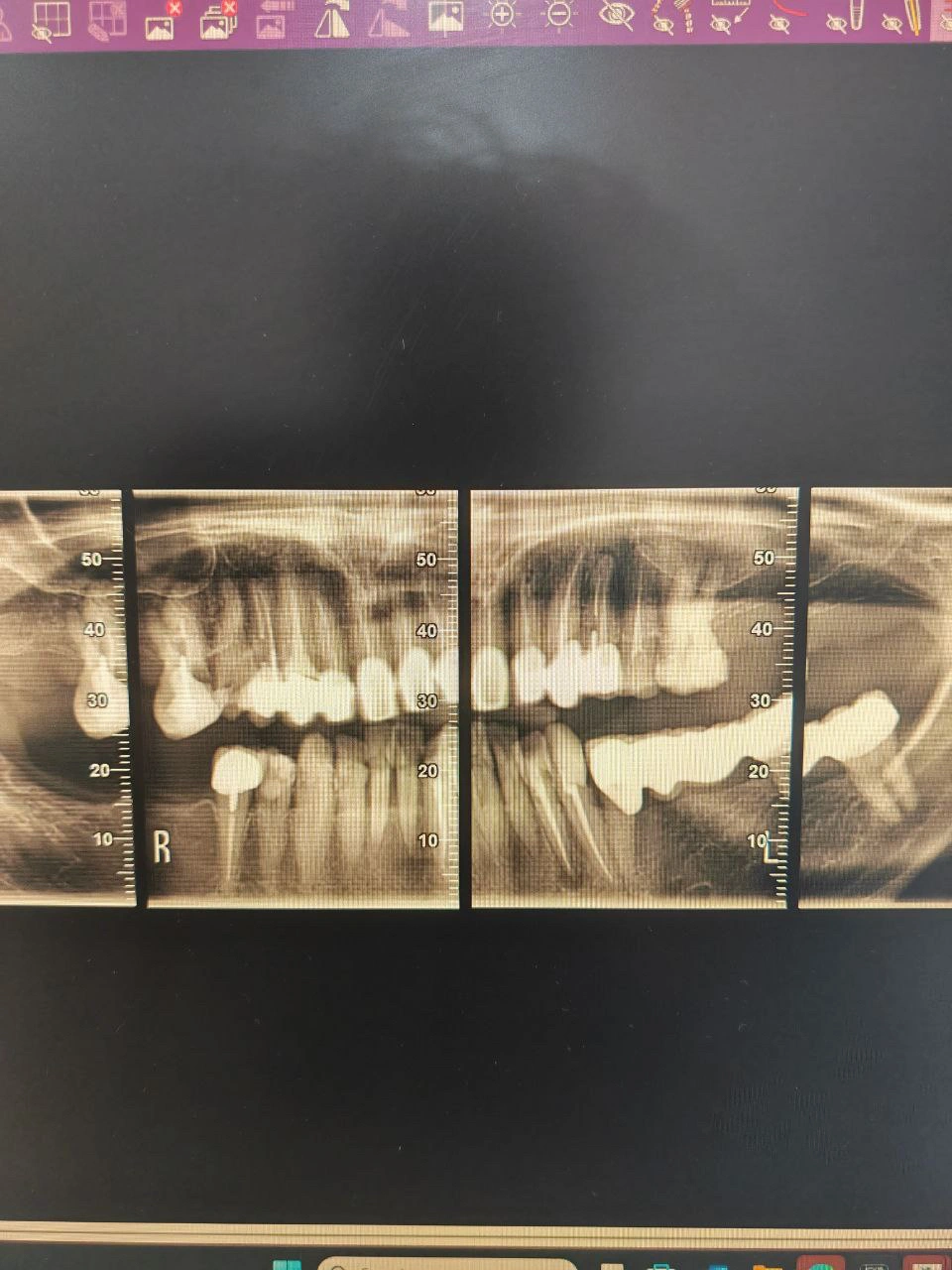
Pre-treatment sectional radiograph - showing multiple missing teeth, previous restorations, and existing bone structure before rehabilitation.
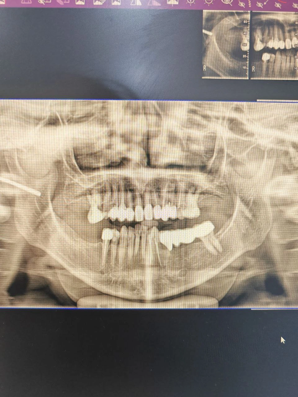
Pre-treatment panoramic radiograph - displaying dentition condition, bone levels, and distribution of existing prosthetics prior to intervention.
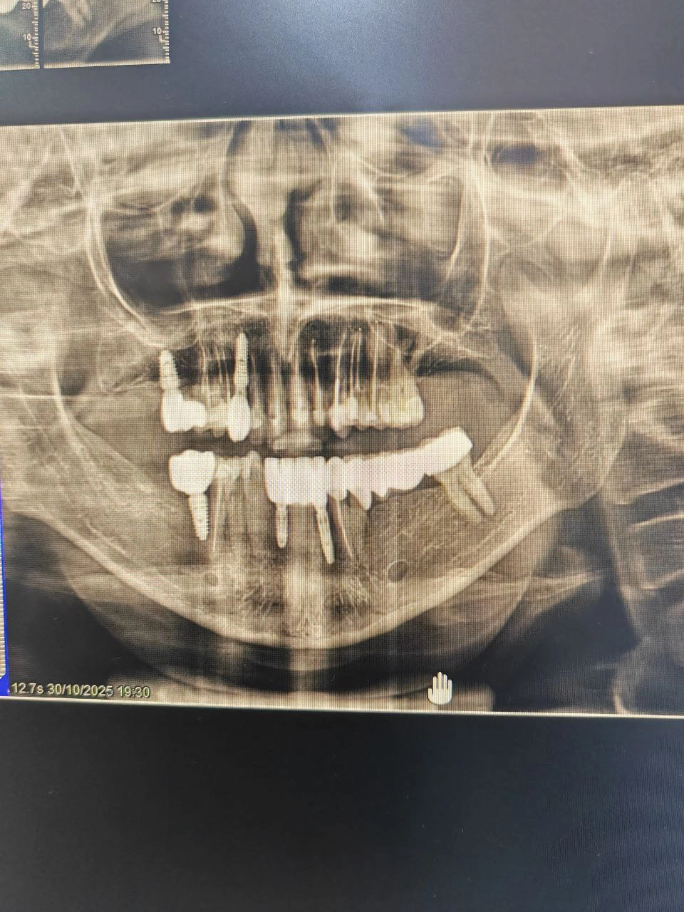
Post-treatment panoramic radiograph - showing successful placement of dental implants and full-arch prosthetic restoration.
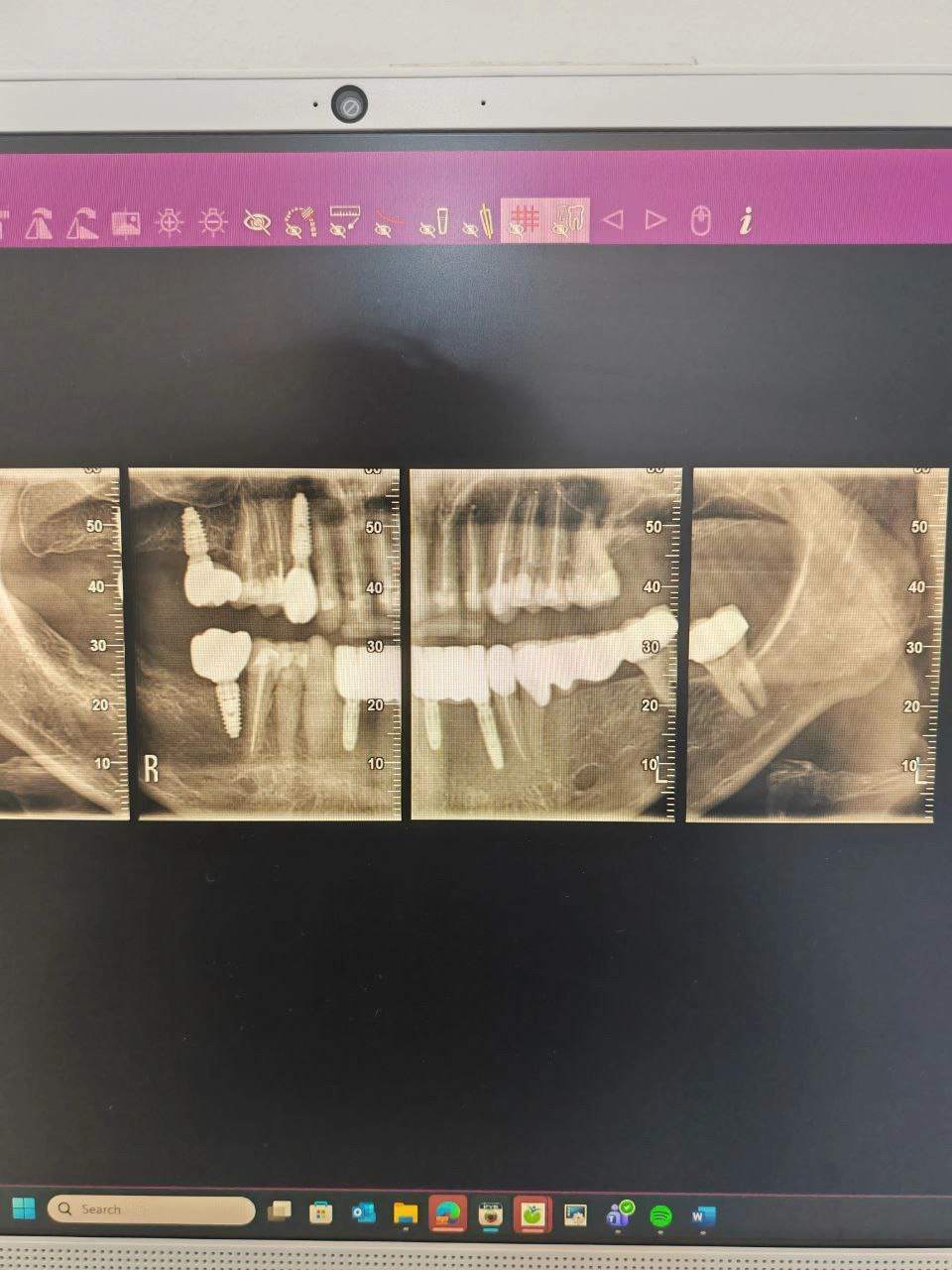
Post-treatment sectional radiograph- confirming implant angulation, depth, and osseointegration supporting the final prosthesis.
Diagnosis
Radiographic Findings (Panoramic & Sectional Views):
Multiple missing teeth in both arches.
Several existing implants with stable bone levels.
Residual roots and failing restorations on premolars and molars.
Evidence of mild generalized bone loss around certain implants.
Clinical Findings:
Reduced masticatory efficiency.
Uneven occlusal plane.
Moderate plaque accumulation on existing prosthetics.
Mild inflammation of peri-implant tissues.
Diagnosis:
Partial edentulism with compromised occlusion and deteriorated prosthetic work.
Indicated for full-mouth prosthetic rehabilitation supported by implants.
Treatment Objectives
Restore full dental function through implant-supported prostheses.
Re-establish vertical dimension and occlusal balance.
Improve aesthetics with natural-looking restorations.
Maintain implant health through proper hygiene and periodic maintenance.
Treatment Plan
Step 1: Full-mouth evaluation and radiographic planning (CBCT + panoramic).
Step 2: Extraction of non-restorable teeth.
Step 3: Placement of new dental implants in edentulous areas.
Step 4: Healing phase and soft tissue management.
Step 5: Fabrication of fixed hybrid prostheses / implant-supported bridges.
Step 6: Occlusal adjustments and follow-up maintenance.
Clinical Procedure Summary
Radiographic Analysis:
Panoramic and sectional radiographs were used to evaluate bone levels, implant angulation, and sinus proximity.Implant Placement: (Done by Implantologist)
Titanium implants placed under guided protocol. Healing abutments installed after osseointegration.Prosthetic Phase:
Impression taken using open-tray technique.
Screw-retained bridges fabricated and torqued to recommended values.Postoperative Care:
Hygiene instructions given and follow-ups scheduled every 3 months.
Results
Stable occlusion restored.
Excellent implant integration and bone support.
Aesthetic and functional outcomes achieved with improved mastication.
Patient reported significant improvement in chewing comfort and smile confidence.
Discussion
This case illustrates the complexity of managing full-mouth rehabilitation in partially edentulous patients.
Success depends on precise implant positioning, occlusal harmony, and patient compliance with maintenance protocols.
Reflection / Learning
Radiographic planning is crucial for long-term implant success.
Interdisciplinary collaboration between oral surgeon and prosthodontist enhances predictability.
Patient motivation and hygiene maintenance determine long-term stability.