Root canal treatment for teeth #4,5,12
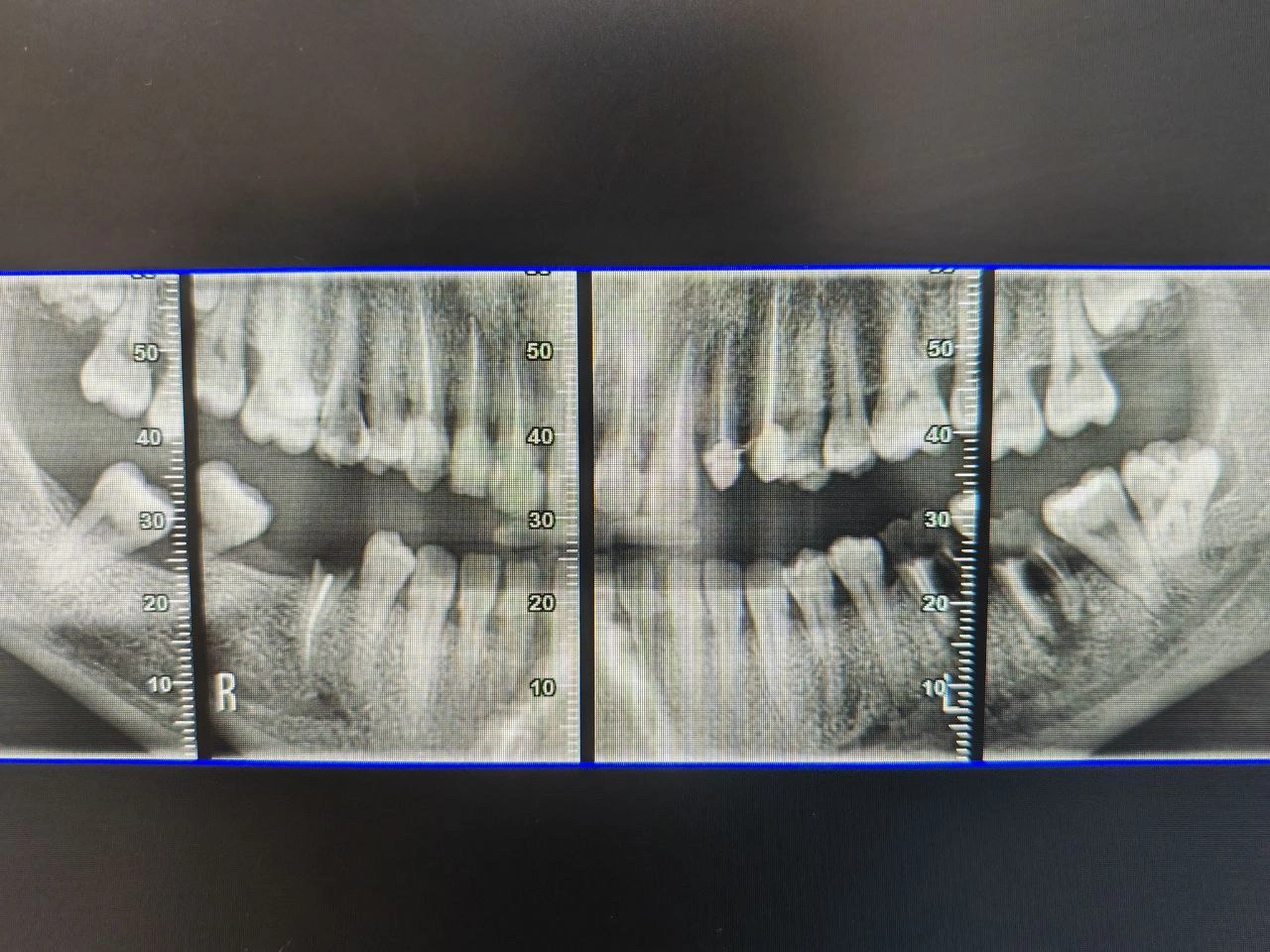
Impacted mandibular third molars bilaterally, with the right molar in close proximity to the inferior alveolar canal.
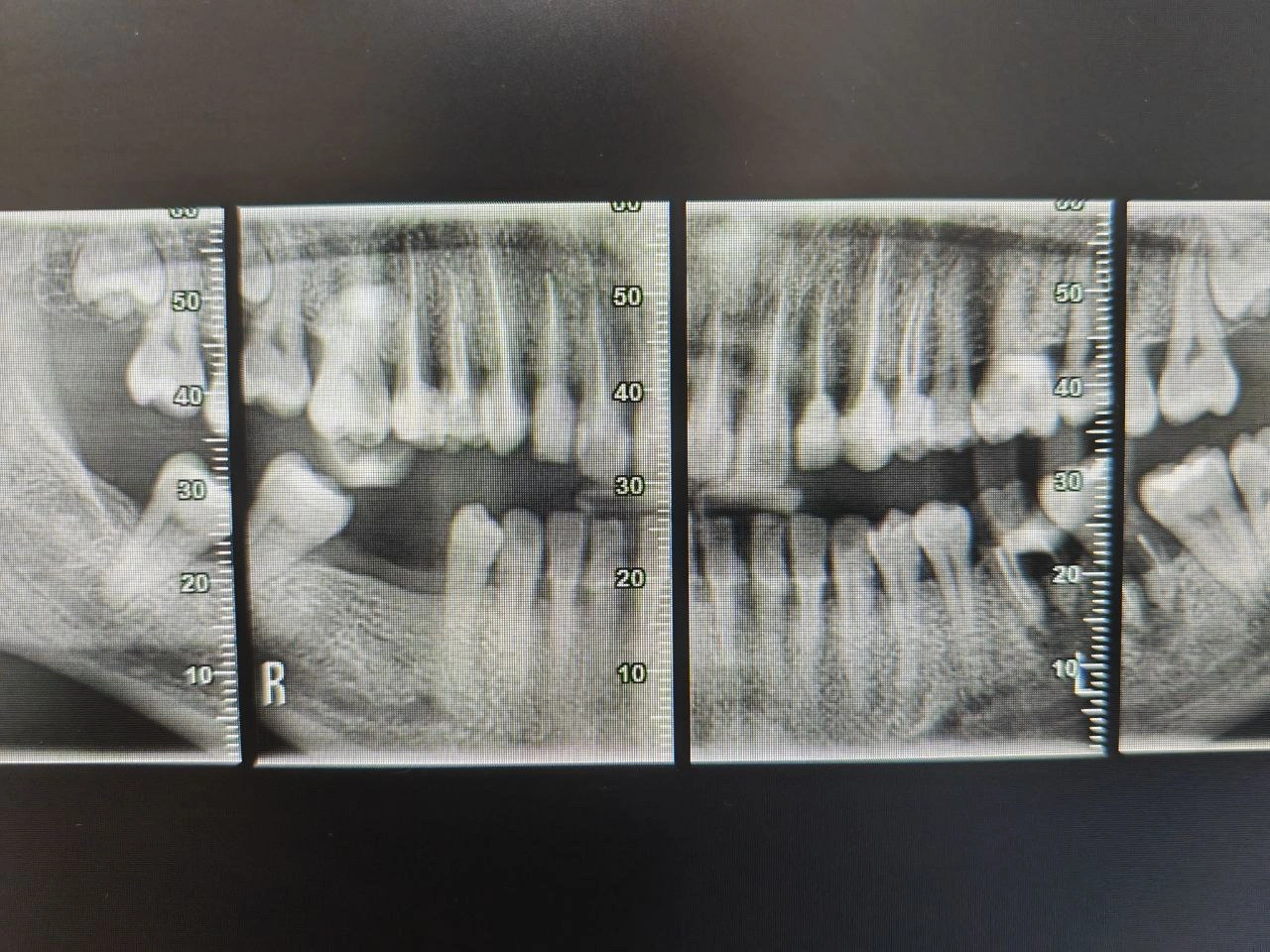
After surgical extraction of the right mandibular third molar, showing clear socket outline and intact canal margins.
Diagnosis
Radiographic Findings:
Carious lesions extending to the pulp of teeth #4, #5, and #12 (irreversible pulpitis).
Clinical Findings:
Mild swelling of the right retromolar area.
Tenderness on palpation; limited mouth opening.
Gingival operculum partially covering the third molar crown.
Diagnosis:
Mesioangular impaction of mandibular third molar associated with pericoronitis.
Treatment Objectives
Eliminate pulpal infection and remove inflamed pulp tissue.
Relieve pain and prevent spread of infection.
Preserve the natural teeth in function and aesthetics.
Achieve a hermetic seal and prevent reinfection.
Restore teeth with durable post-endodontic restorations.
Treatment Plan
Pre-operative phase:
Clinical and radiographic evaluation, diagnosis confirmation, and informed consent.
Endodontic phase:
Local anesthesia.
Access cavity preparation.
Working length determination with apex locator and radiograph.
Cleaning and shaping using rotary or hand files with NaOCl and EDTA irrigation.
Obturation with gutta-percha and sealer (Vertical technique).
Temporary.
Restorative phase:
Core build-up to restore lost tooth structure.
Post-operative care:
Radiographic and clinical follow-up for periapical healing.
Results
Teeth #4, #5, and #12 became asymptomatic with complete relief of pain.
Radiographs showed adequate obturation and periapical healing.
No post-operative sensitivity or signs of reinfection were noted at review.
Reflection / Learning
Reinforced the importance of accurate diagnosis and aseptic isolation during endodontic therapy.
Improved confidence in multi-tooth RCT planning and time management.
Gained better understanding of communication with patients about treatment phases and expectations.