Aesthetic Rehabilitation with Porcelain Veneers and Crowns

Pre-Treatment (Retracted Intraoral View) — highlights severe staining and surface irregularities on upper anterior teeth.
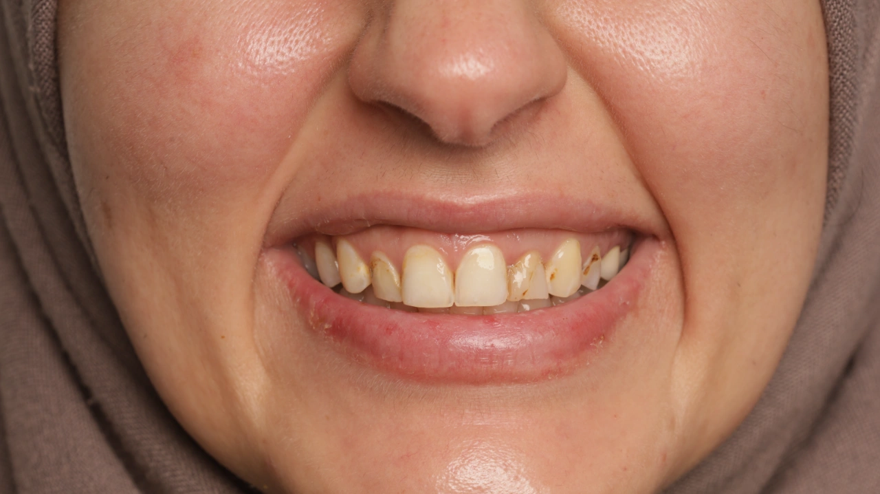
Pre-Treatment (Frontal View) — showing intrinsic and extrinsic discoloration, enamel wear, and irregular incisal edges.
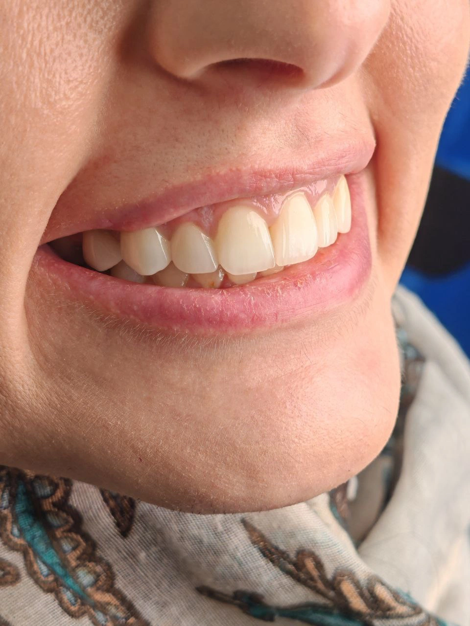
Post-Treatment (Right Smile View) — natural, bright, and symmetrical appearance following veneer placement.
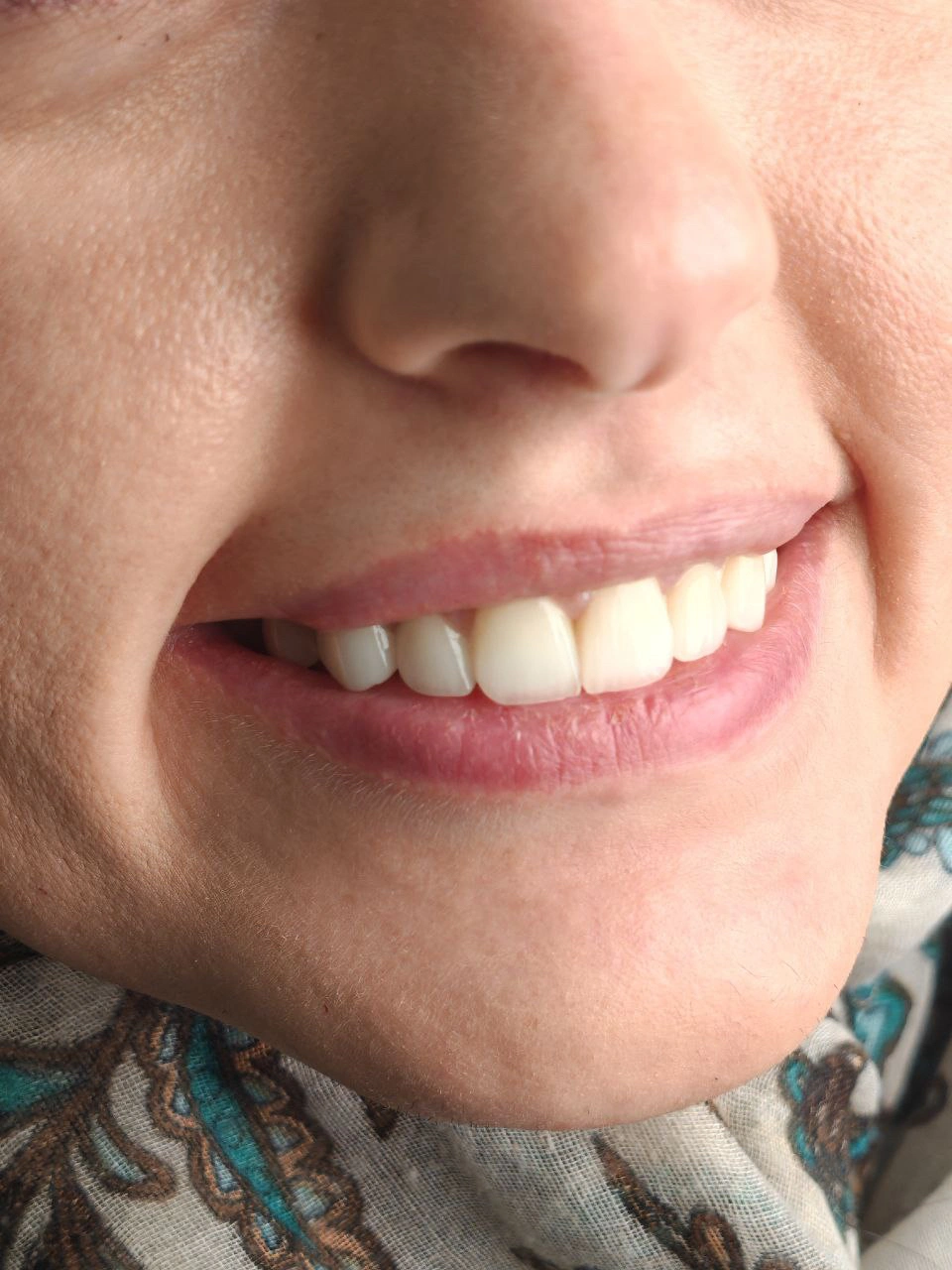
Post-Treatment — harmonious shade match and enhanced gingival aesthetics.
Diagnosis
Dental Findings:
Extrinsic and intrinsic discoloration of anterior teeth.
Mild enamel attrition and irregular incisal edges.
Surface staining likely from dietary and environmental factors.
Slight gingival asymmetry, but healthy periodontium overall.
Diagnosis:
Aesthetic disharmony caused by enamel discoloration and surface irregularities.
Indicated for cosmetic rehabilitation with porcelain veneers.
Treatment Objectives
Eliminate discoloration and restore uniform shade across anterior teeth.
Correct shape irregularities to enhance smile symmetry.
Preserve enamel through minimally invasive veneer preparation.
Achieve a natural, translucent, and aesthetically pleasing smile.
Treatment Plan
Step 1: Preoperative photo documentation and shade analysis.
Step 2: Smile design using digital mock-up to visualize the Final outcome.
Step 3: Minimal tooth preparation (≈0.3–0.5 mm) confined to enamel.
Step 4: Impression with high-precision material / intraoral scanning.
Step 5: Fabrication of E.max porcelain veneers/crowns.
Step 6: Try-in and cementation using light-cured resin cement.
Step 7: Final finishing and occlusal adjustments.
Clinical Procedure
Preoperative Evaluation:
Documented initial discoloration, surface stains, and enamel wear.Tooth Preparation:
Minimal reduction, maintaining enamel integrity for optimal bonding.Isolation and Cementation:
Veneers etched with hydrofluoric acid and salinized; enamel etched with 37% phosphoric acid.
Bonding achieved with dual-cure resin cement.Finishing:
Margins polished and occlusion checked in centric and lateral movements.Postoperative Photos:
Captured to evaluate colour match, symmetry, and gingival harmony.
Results
Achieved a harmonious smile line with excellent colour uniformity and natural translucency.
The patient reported increased confidence and satisfaction with her new smile.
Gingival contour and enamel luster appeared healthy and balanced.
Postoperative photos clearly demonstrate enhanced shade, surface texture, and smile symmetry.
Discussion
This case highlights the power of minimally invasive porcelain veneer therapy in restoring esthetics while preserving natural tooth structure.
Proper shade selection, smile design, and adhesive protocols are crucial for achieving long-term success and natural results.
Reflection / Learning
Comprehensive preoperative planning ensures predictable results.
Careful enamel preservation enhances bonding strength and veneer longevity.
Patient education and maintenance (avoiding staining foods, regular hygiene) are key for durability.