RCT #3
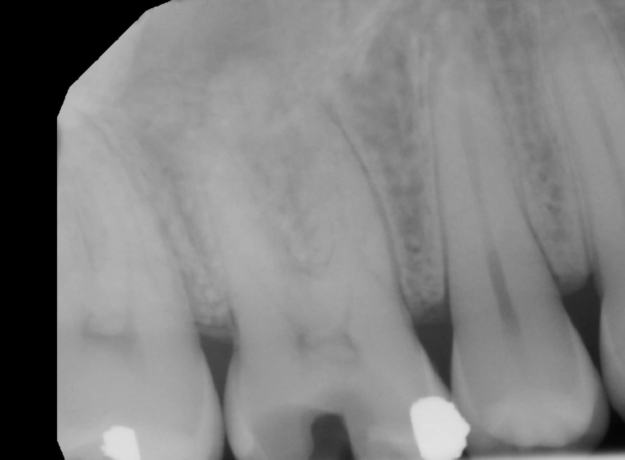
Pre-Operative
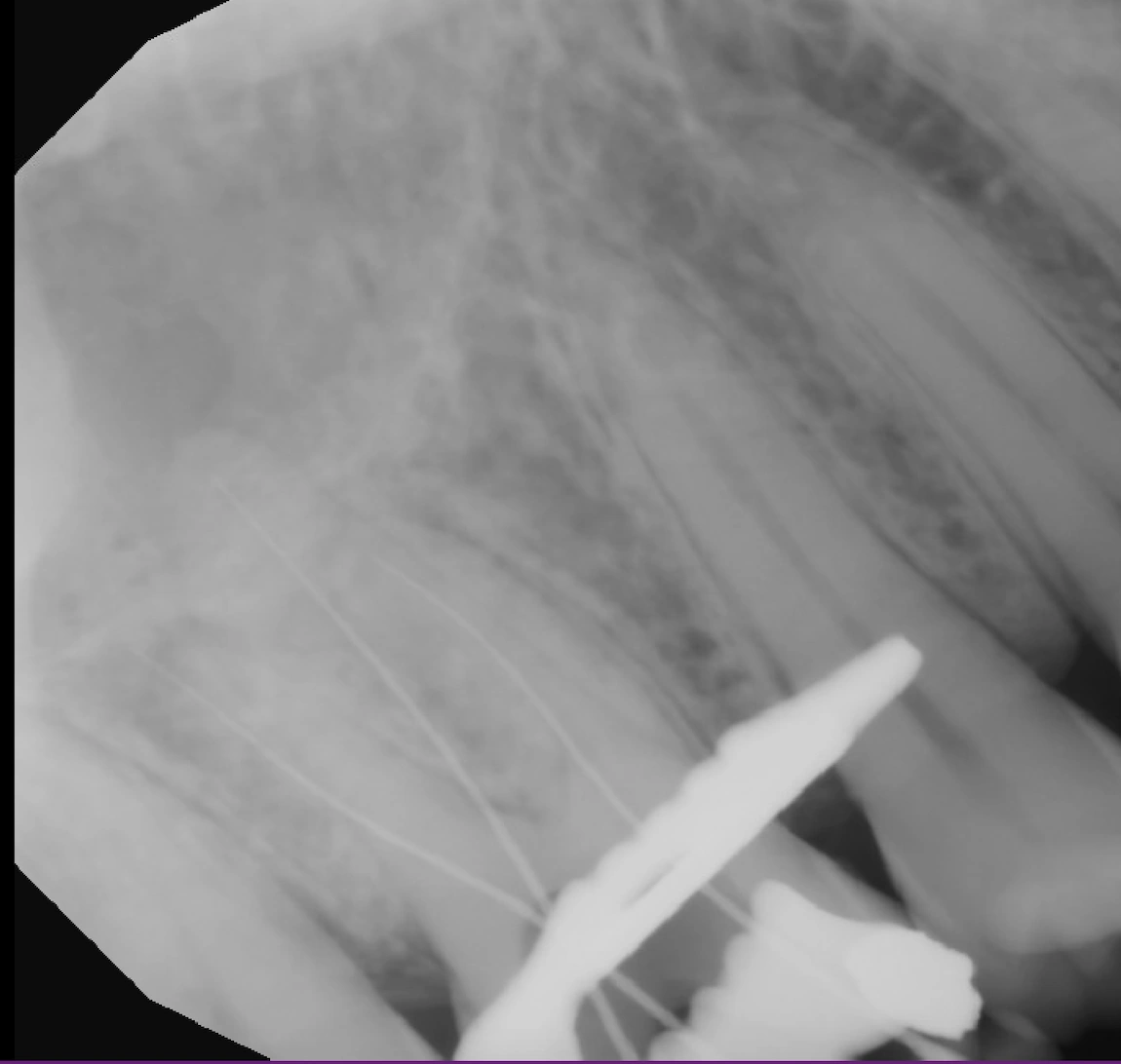
Working Length x-ray
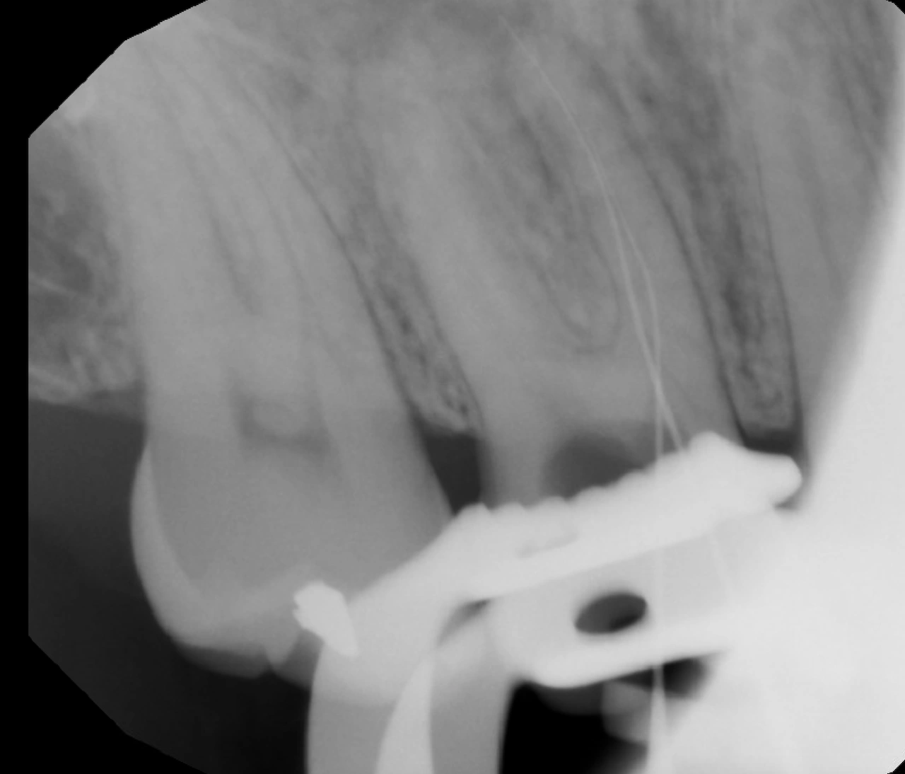
Working Length x-ray of MB & MB2 to visualize anatomy
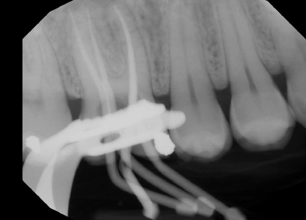
Master Cone x-ray
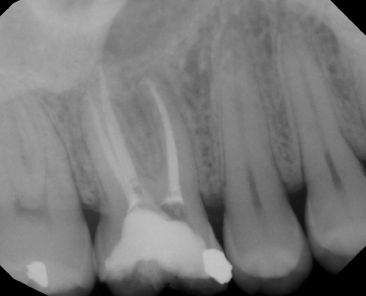
Obturation Complete
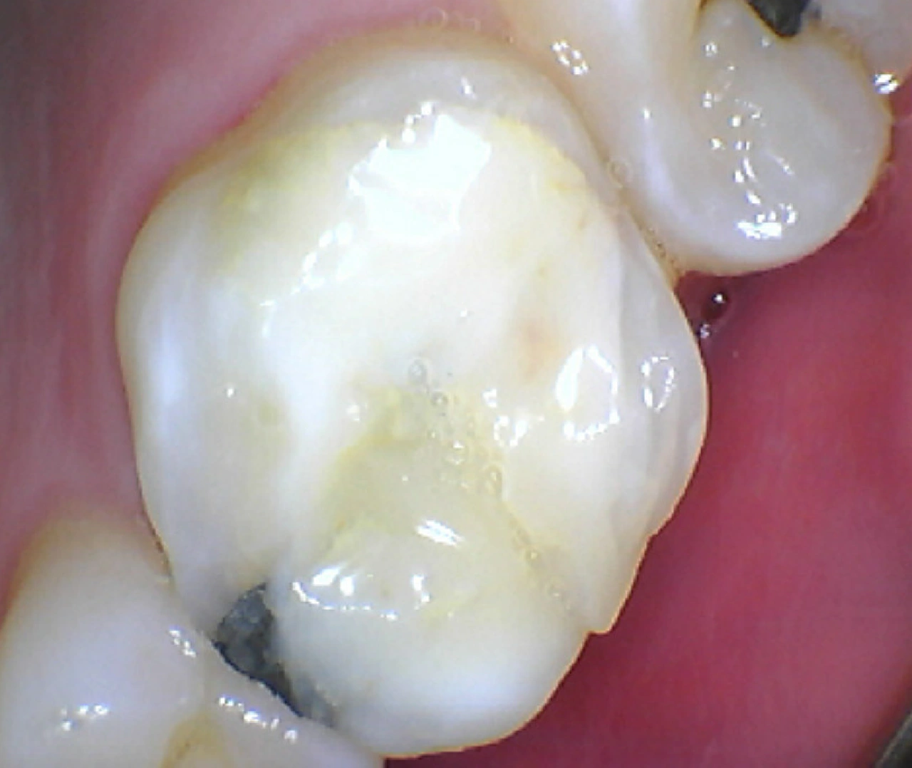
Next Visit Post-Op: post and core build up
Tooth #3
The patient presented with a chief complaint of pain on biting and intermittent discomfort. Clinical examination revealed tenderness to percussion and palpation. Periodontal probing depths were within normal limits. Pulp vitality testing with cold and EPT yielded no response. Radiographic evaluation showed widening of the lamina dura around the mesial root and a periapical radiolucency associated with the palatal root.
A diagnosis of necrotic pulp with symptomatic apical periodontitis was made. Treatment was completed in 2 visits, first visit was an emergency pulpectomy, 3 canals were located then. The second visit, four canals were located as MB2 was found. A separate working length radiograph of the mesial canals was taken to better visualize the canal anatomy and confirm working length. The canals were instrumented using a combination of manual files and the WaveOne Gold system (Primary/Red). After thorough irrigation and cleaning, the canals were obturated with matching WaveOne GP cones.
Following completion of root canal treatment, the tooth required a post and core due to significant loss of coronal structure. The tooth is currently undergoing definitive restoration with a full coverage crown.