RCT #19
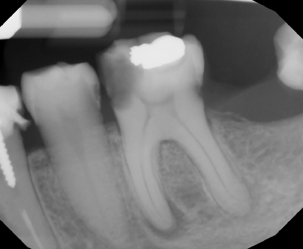
Pre-Operative PA
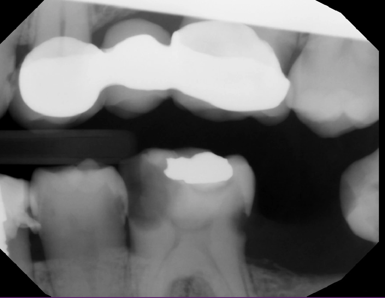
Pre-Operative BW
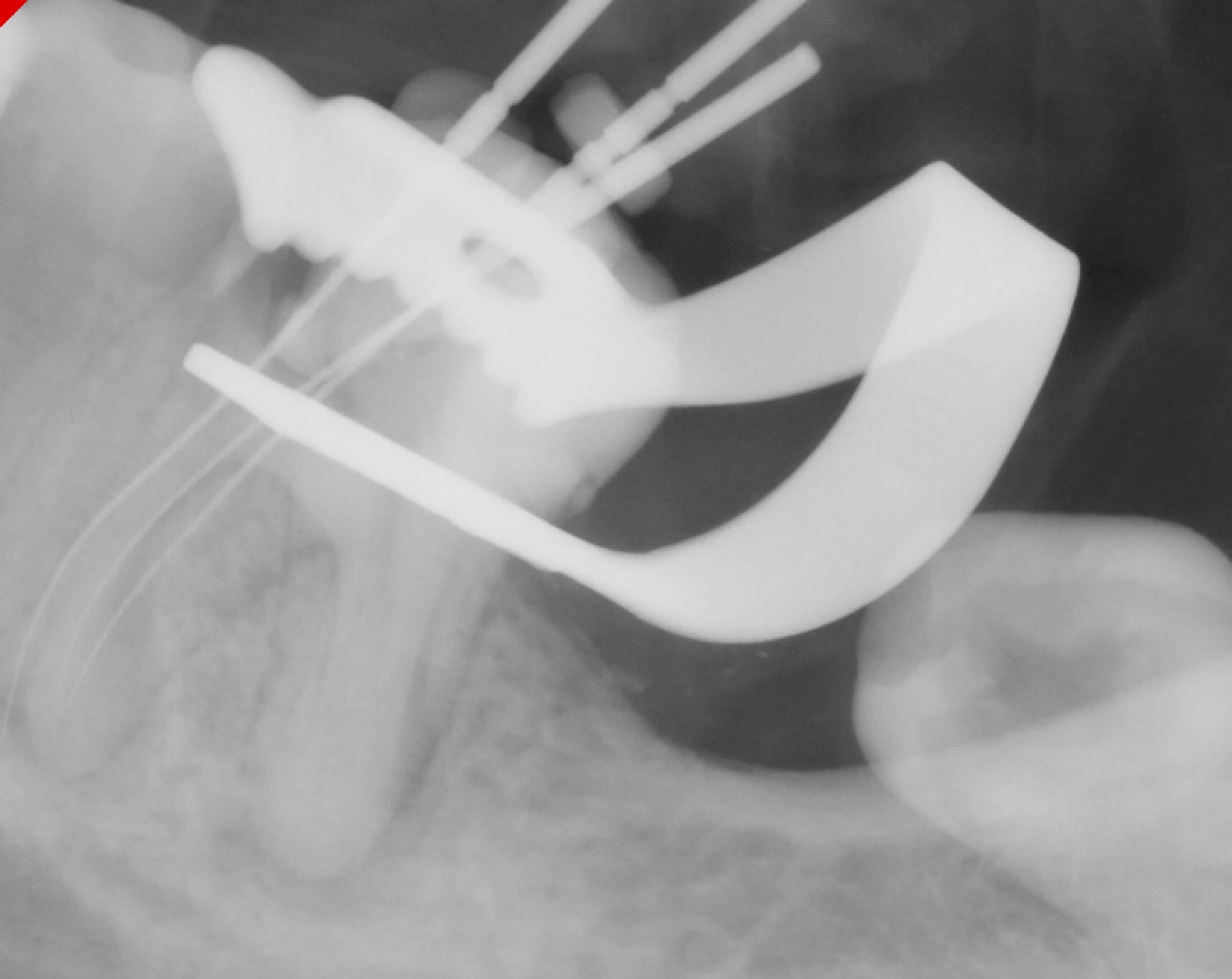
3 Mesial Canals WL, Mid-mesial joing ML
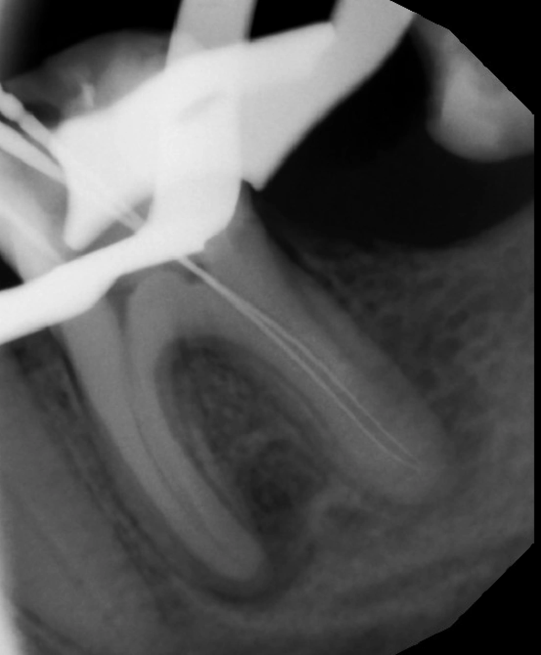
Distal Canals WL (added 1mm)
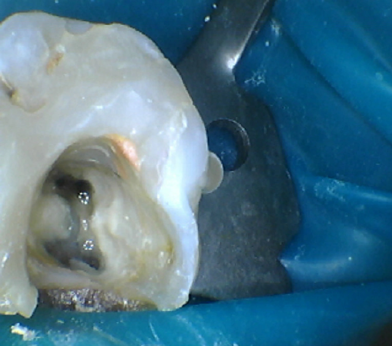
Orifice Widening For Better Instrumentation
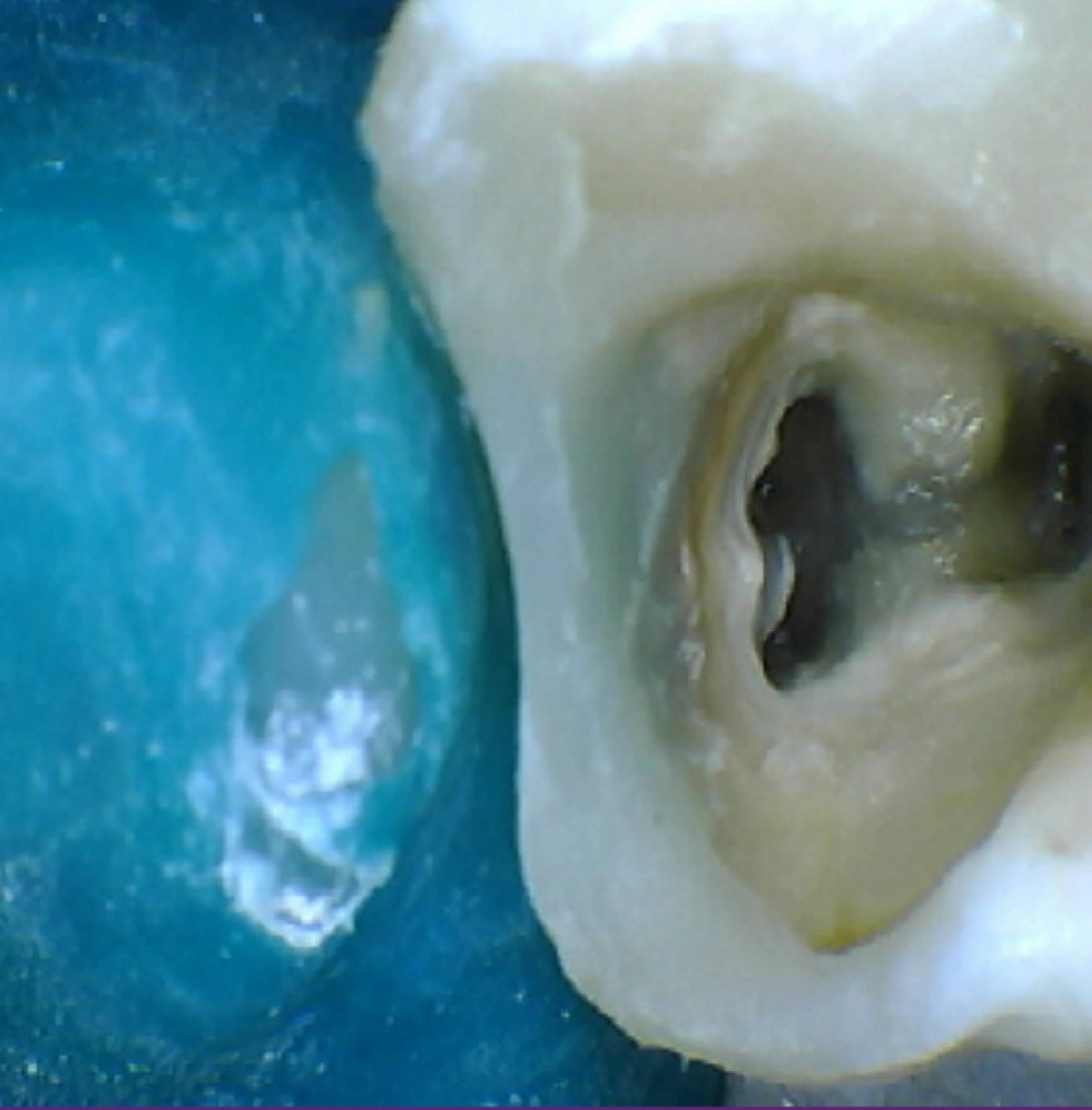
Mesial Wall Build Up to Achieve Seal
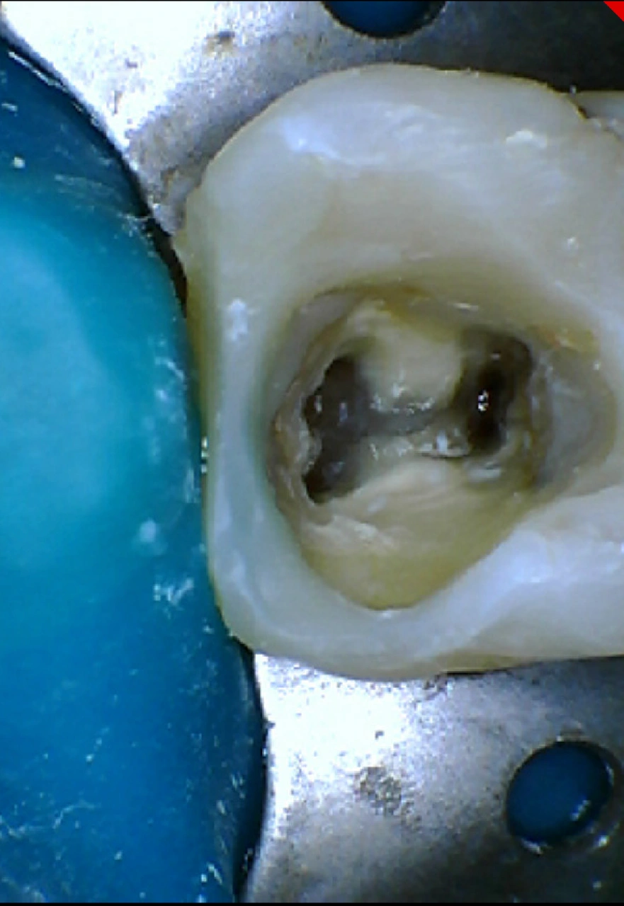
Second Visit
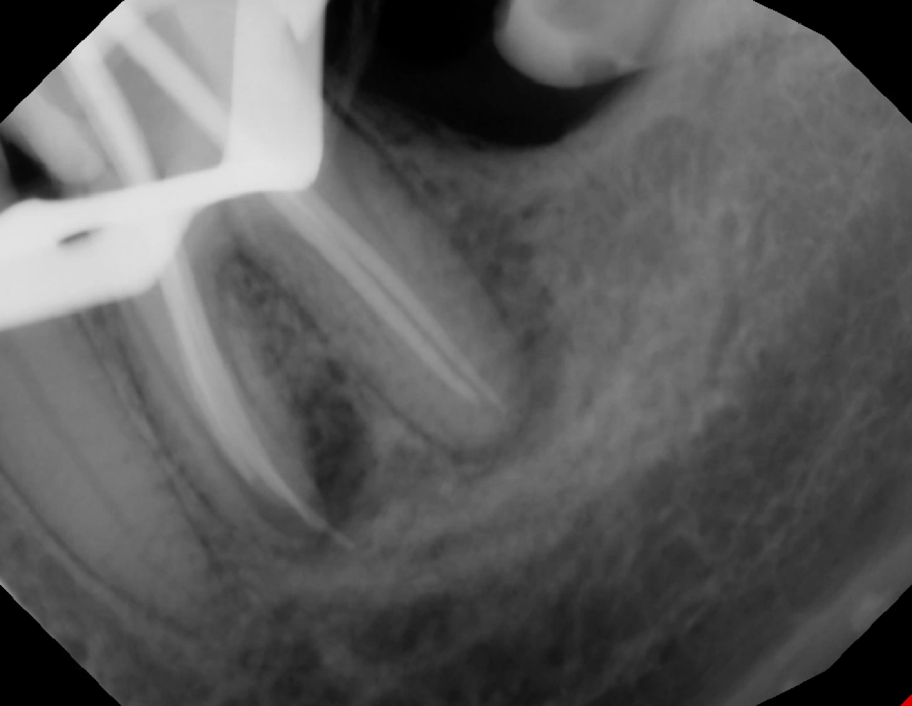
Master Cone, ML canal is past the apex, DB canal is 1mm short
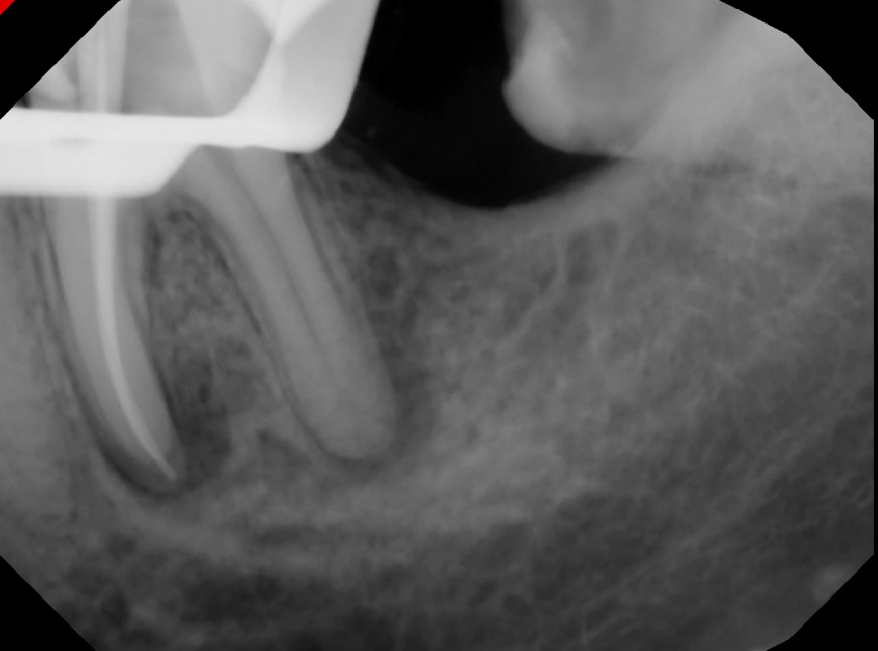
Master Cone, ML and Midmesial Canals
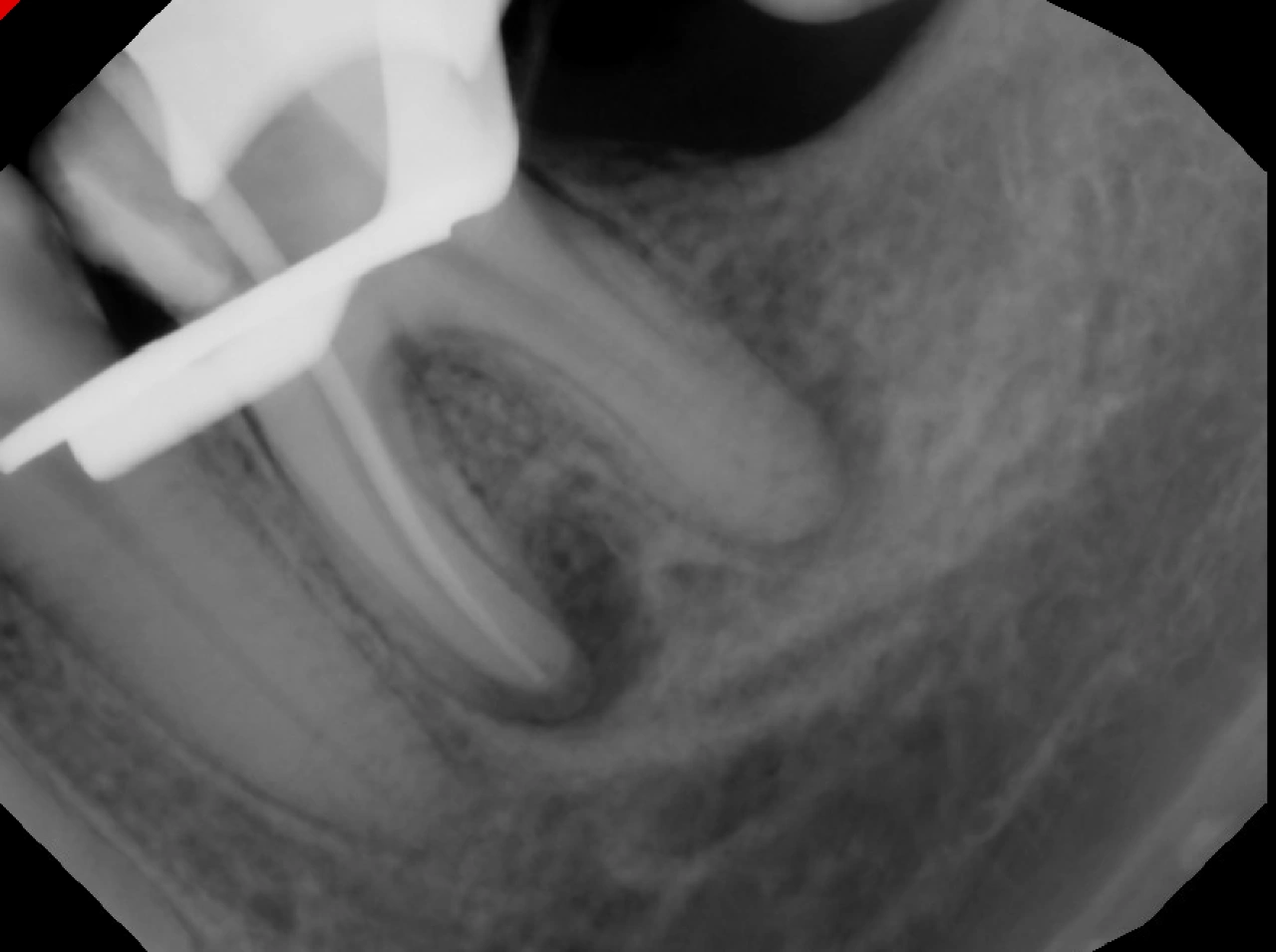
Master Cone, MB Canal Taken Separately
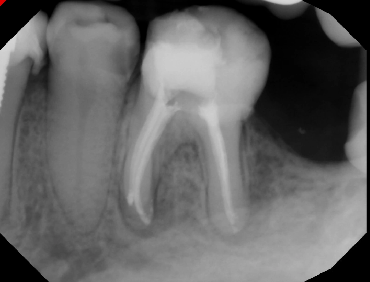
Obturation Complete
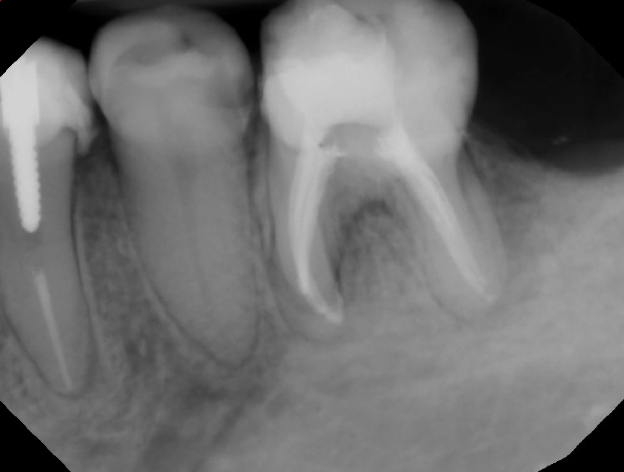
Mesial Shift
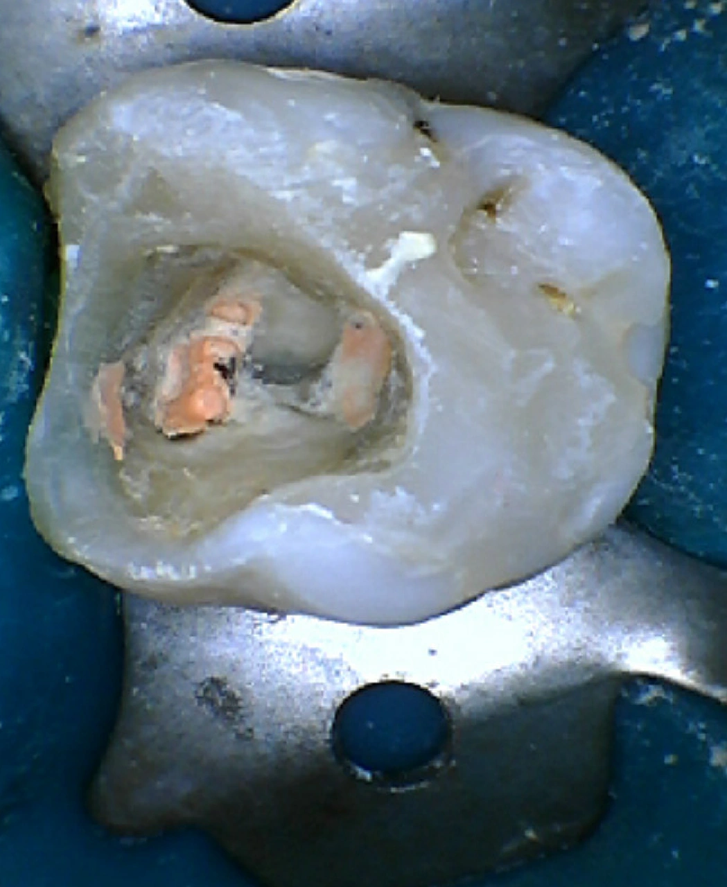
Obturation Complete, Excess GP was cleaned off from the chamber
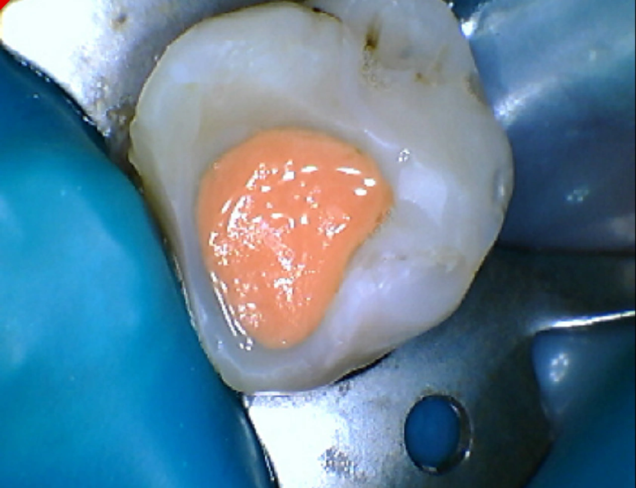
Temporary Filling (Fuji Pink)
A 35-year-old female was referred by a colleague for treatment of tooth #19. Clinical and radiographic evaluation revealed a necrotic pulp with chronic apical abscess.
At the first visit, an emergency pulpectomy was performed. Four canals were initially identified and instrumented. The second visit was completed in the postgraduate endodontic clinic (7W) under the dental operating microscope. During this visit, a mid-mesial canal was located, traced, and working length radiographs were taken for all mesial canals. The mid-mesial canal joined the mesiolingual canal. Two distal canals were also identified and prepared. To ensure an adequate coronal seal, a mesial wall build-up was performed using flowable composite prior to obturation.
At the next visit, all canals were cleaned and shaped, waveone red was used for ML and DL canals, and waveone small was used for MB, Midmesial and DB.
Obturation was completed and tooth is now temporized to be restored by primary provider.